Nanoparticle Surface Protein Analysis
NanoFCM
Characterize nanoparticles and extracellular vesicles (EVs) from almost any fluid and biofluid - from plasma to tissue culture media
Nano-flow cytometry is the latest development for protein analysis on nanoparticles. Using light scattering and labelled fluorescent antibodies, our analysis allows direct, single-particle physical and bio-chemical measurements of nano-sized entities on a single platform.
- Detection of submicron particles, including exosomes and lipoproteins (Fig. 1.)
- Up to 3 fluorescent signals for multiparameter sub-population analysis
- Low sample volume requirement
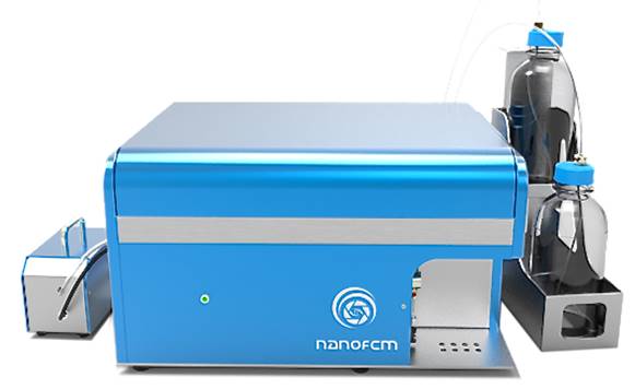
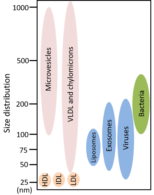
Fig. 1. Detectable range of EVs
(30nm – 2000nm)
NanoFCM Case Study
Using fluorescence and measurements of particle size and concentration to determine breast cancer HER2 status
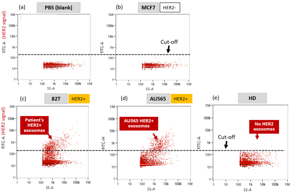
HER2 is a growth-promoting protein found on 15-20% of breast cancer cells. These cells tend to grow and spread faster than normal breast cells. In the following analysis, HER2 antibodies were labeled exosomes from MCF7 cell line (HER2-Negative), 82T primary cell line (HER2-Positive metastatic site), AU565 cell line (HER2-Positive), and healthy donor (HD), were tested with PBS (blank) as control. The figure below shows HER2-Positive signals above the cut-off for 82T and AU565. The other signals below cut-off indicate HER2-Negative.
Publication Library
Read about Nano-flow cytometry (NanoFCM) applications in industry and other research uses in these published papers.
Sample Reports
Sample Preparation Instructions
Contact us at info@reliance-bio.com (general inquiries)
FAQs
Click here for FAQs